| Right atrial enlargement | |
|---|---|
 | |
| Right atrial enlargement (P pulmonale) | |
| Causes | Pulmonary Hypertension |
| Diagnostic method | electrocardiogram |
Right atrial enlargement (RAE) is a form of cardiomegaly, or heart enlargement. It can broadly be classified as either right atrial hypertrophy (RAH), overgrowth, or dilation, like an expanding balloon. Common causes include pulmonary hypertension, which can be the primary defect leading to RAE, or pulmonary hypertension secondary to tricuspid stenosis; pulmonary stenosis or Tetralogy of Fallot i.e. congenital diseases; chronic lung disease, such as cor pulmonale. Other recognised causes are: right ventricular failure, tricuspid regurgitation, and atrial septal defect.[1] Right atrial enlargement (RAE) is clinically significant due to its prevalence in diagnosing supraventricular arrhythmias. Further, early diagnosis using risk factors like RAE may decrease mortality because patients with RAE are at 9x more risk of arrhythmias and other cardiac conditions compared to their healthy counterparts (Waligóra et al.). [2]
Diagnosis
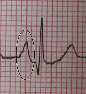
Right Atrial Enlargement (RAE) increases the p wave, representing atrial depolarization, on an ECG to an amplitude > 2.5mm in lead II, an abnormality referred to as p-pulmonale, likely due to weakened right atrial myocardium close to the Sinoatrial (SA) node (Limongelli et al., 2017). [3]
ECG criteria for RAE: P wave amplitude in lead II > 2.5 mm and upward deflection of the P wave in lead V1 > 1.5 mm in amplitude.[4]
Large "a" waves on the JVP waveform can also aid in diagnosis.
References
- ↑ "Size of the right atrium and associated structures". 123 sonography.com. Medical University of Vienna. 2012-08-20. Retrieved 13 December 2016.
- ↑ https://www.sciencedirect.com/science/article/abs/pii/S0147956317302169?casa_token=k8s9x4j-BtQAAAAA:G0ESkEIaC7PjxjB_ZyCaWfIezSP569GKk5ju94AFu5P1at--Veif-GM6vQVF6Wqv_hXcAs6Q6Q
- ↑ doi:10.2459/JCM.0000000000000361
- ↑ Surawicz, Borys; Childers, Rory; Deal, Barbara J.; Gettes, Leonard S. (2009). "AHA/ACCF/HRS Recommendations for the Standardization and Interpretation of the Electrocardiogram". Circulation. 119 (10). doi:10.1161/circulationaha.108.191095.