| ATP-cone | |||||||||
|---|---|---|---|---|---|---|---|---|---|
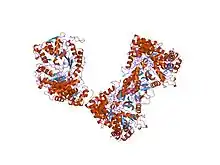 ribonucleotide reductase e441q mutant r1 protein from escherichia coli | |||||||||
| Identifiers | |||||||||
| Symbol | ATP-cone | ||||||||
| Pfam | PF03477 | ||||||||
| InterPro | IPR005144 | ||||||||
| SCOP2 | 7r1r / SCOPe / SUPFAM | ||||||||
| |||||||||
In molecular biology, the ATP-cone is an evolutionarily mobile, ATP-binding regulatory domain which is found in a variety of proteins including ribonucleotide reductases, phosphoglycerate kinases and transcriptional regulators.[1]
In ribonucleotide reductase protein R1 from Escherichia coli this domain is located at the N terminus, and is composed mostly of helices.[2] It forms part of the allosteric effector region and contains the general allosteric activity site in a cleft located at the tip of the N-terminal region.[3] This site binds either ATP (activating) or dATP (inhibitory), with the base bound in a hydrophobic pocket and the phosphates bound to basic residues. Substrate binding to this site is thought to affect enzyme activity by altering the relative positions of the two subunits of ribonucleotide reductase.
References
- ↑ Aravind L, Wolf YI, Koonin EV (April 2000). "The ATP-cone: an evolutionarily mobile, ATP-binding regulatory domain". J. Mol. Microbiol. Biotechnol. 2 (2): 191–4. PMID 10939243.
- ↑ Uhlin U, Eklund H (August 1994). "Structure of ribonucleotide reductase protein R1". Nature. 370 (6490): 533–9. doi:10.1038/370533a0. PMID 8052308. S2CID 8940689.
- ↑ Eriksson M, Uhlin U, Ramaswamy S, Ekberg M, Regnstrom K, Sjoberg BM, Eklund H (August 1997). "Binding of allosteric effectors to ribonucleotide reductase protein R1: reduction of active-site cysteines promotes substrate binding". Structure. 5 (8): 1077–92. doi:10.1016/S0969-2126(97)00259-1. PMID 9309223.