< Radiation Oncology < Thymoma
|
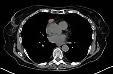
Thymoma Overview
Epidemiology
- Arises from thymus epithelial cells
- Mostly considered indolent, but does have potential for local invasion, pleural dissemination, and distant metastases
- Malignant thymoma (see below, but in the SEER study defined as microscopic or macroscopic evidence of invasion) is rare; there were 849 cases in US SEER database between 1973-1998 (incidence 0.15/100,000)
- Age typically 40-60 years; though highest incidence in 70's
- M:F ~1:1
- 20% of primary mediastinal neoplasms in adults
- Risk factors:
- Myasthenia gravis in 35-50% (conversely, 75% patients with MG have thymus abnormalities, of these 85% have hyperplasia and 15% have thymoma)
- Red cell aplasia in 5%
- Hypogammaglobulinemia in 5%
- Ionizing radiation
- Clinical presentation:
- ~1/3 asymptomatic, found incidentally on CXR
- ~1/3 local symptoms (chest pain, neck mass, SVC syndrome
- ~1/3 concurrent diagnosis with myasthenia gravis
- SEER; 2003 (1973-1998) PMID 12712448 -- "Malignant thymoma in the United States: demographic patterns in incidence and associations with subsequent malignancies." (Engels EA, Int J Cancer. 2003 Jul 1;105(4):546-51.)
- Population study. Malignant thymoma (vs. benign thymoma and possibly vs. thymic carcinoma) reviewed. 849 cases (0.15/100,000). Male slightly more than females. Highest incidence in Pacific Islanders (0.49/100,000)
- Follow-up malignancies: Sarcoma 11.1x, NHL 4.7x (all after RT), GI cancers 1.8x
Anatomy
- Mediastinum borders
- Superior: thoracic inlet
- Inferior: diaphragm
- Anterior: sternum
- Posterior: vertebral column
- Lateral: parietal pleural
- Thymoma typically presents in the anterior/superior mediastinum
- Spread is mainly local invasion; pleural mets may occur in more advanced disease
Histology
- Thymus is unique among human organs because its "normal" appearance varies considerably depending on age of the patient
- At birth, weight is 10-35gm
- Functional maturity during childhood/adolescence, with weight 20-50gm
- In adults, progressive atrophy of thymic tissue, and replacement with fat
- In elderly, weight 5-15gm
- Two predominant cell types in normal thymus
- Thymic epithelial cells
- Lymphocytes (T-cell lineage)
- Thymus anatomy
- Two fused lobes, comprising of multiple lobules
- Each lobule has an outer cortex and inner medulla
- Thymic epithelial tumors arise from thymic epithelial cells, but due to the role of thymus in lymphocyte maturating, they may predominately contain lymphocytes
- There are multiple classifications of thymic epithelial tumors, making things confusing for clinicians
- The WHO 1999/2004 morphologic classification is the most commonly accepted, and has been shown to have correlation with clinical behavior and outcome
- A proposal has been made for a more simplified classification parallel to other carcinomas (thymoma, atypical thymoma, thymic carcinoma)
Bernatz Classification System for Thymoma (1961)
- Spindle Cell (favorable outcome)
- Lymphocytic
- Epithelial (aggressive)
- Mixed (aggressive)
WHO Classification of Thymic Epithelial Tumors (1999/2004) - based on shape and lymphocyte/epithelial ratio
- Thymoma (Type A, B, and AB)
- Type A - Spindle/ovoid shape, homogeneous neoplastic cells, few lymphocytes; equivalent to "spindle cell tumor"
- Type AB - Mix of Type A and Type B, with foci rich in lymphocytes
- Type B - Round/polygonal shape, with varying proportion of lymphocytes/epithelial cells, and increasing degree of atypia
- Type B1 - Resembles normal thymus, with areas resembling both cortex and medulla. Few neoplastic epithelial cells, predominance of lymphocytes; equivalent to "lymphocyte-rich tumor"
- Type B2 - Increased number of epithelial thymic cells, among populations of lymphocytes; equivalent to "mixed tumors"
- Type B3 - Predominately neoplastic epithelial cells, more atypia, with few lymphocytes
- Thymic carcinoma (Type C)
- Over features of malignancy independent of shape, no immature lymphocytes
- Multiple histologic subtypes
Suster and Moran (1999) - based on degree of differentiation; PMID 16627265
- Thymoma - well differentiated
- Atypical thymoma - moderately differentiated
- Thymic carcinoma - poorly differentiated
- Osaka; 2002 PMID 11857293 -- "The World Health Organization histologic classification system reflects the oncologic behavior of thymoma: a clinical study of 273 patients." (Okumura M, Cancer. 2002 Feb 1;94(3):624-32.)
- Retrospective. 273 with thymoma (no thymic carcinoma).
- Invasive by class: A 11%, AB 42%, B1 47%, B2 69%, B3 85%
- Great vessel invasion by class: A 0%, AB 4%, B1 7%, B2 17%, B3 19%
- 20-year OS: A 100%, AB 87%, B1 91%, B2 59%, B3 36%
- By Masaoka stage: I 89%, II 91%, III 49%, IV 0%
- Multivariate predictors: Masaoka and WHO class; not related R-status, great vessel involvement
- Conclusion: WHO histologic appearance reflects oncologic behavior
Work-Up
- CT scan
- MRI yield over CT minimal
- Serum AFP and b-HCG to rule out germ cell tumors
- UCLA; 2008 -- PMID 18517274 -- "Evidence-based pathology and the pathologic evaluation of thymomas: transcapsular invasion is not a significant prognostic feature." (Gupta R, Arch Pathol Lab Med. 2008 Jun;132(6):926-30.)
- Meta-analysis. 21 retrospective publications, 2451 cases (Stage I 1419, Stage II 1032)
- Outcome: No difference in DFS or OS for Stage I and Stage II
- Conclusion: Evaluation of transcapsular invasion is of no clinical value in tumors that lack invasion of neighboring organs or the pleurra
- MSKCC; 1999 (1949-1993) PMID 9475524 -- "Thymic carcinoma: current staging does not predict prognosis." (Blumberg D, J Thorac Cardiovasc Surg. 1998 Feb;115(2):303-8; discussion 308-9.)
- Retrospective. 43 patients, Masaoka Stage I 7%, Stage II 35%, Stage III 47%, Stage IVA 11%. Well-differentiated 16/43, type II malignant thymomas 27/43
- Outcome: OS 5-years 65%, 10-years 35%; recurrence rate 65% and 75%
- Predictors: multivariate only invasion of innominate vessels, not age, sex, size, or Masaoka stage
- Conclusion: high rate of recurrence; invasion of the innominate vessels poor sign. Masaoka staging not useful for thymic carcinoma, only for thymoma
- Masaoka comment: PMID 10047676
Treatment Overview
- Total thymectomy with en bloc removal of all affected structures is the surgical procedure of choice. Degree of resection appears to affect prognosis (GTR > STR > biopsy alone)
- Patients with Stage I disease and R0 resection do not need adjuvant treatment
- Adjuvant treatment of patients with Stage II disease is controversial; adjuvant RT may be indicated
- Recent pathological meta-analysis (see above, PMID 18517274) suggests that there is no difference in outcome between Stage I and IIa
- If pleural invasion (T2b), should consider RT
- If close (<1mm) surgical margin, can consider RT
- If "higher" grade (e.g. WHO B3), can consider RT
- Adjuvant RT appears indicated with Stage III/IVA or subtotal resection (R1 or R2)
- NCCN Adjuvant Guidelines (2012) - Conventional fractionation (1.8-2Gy per fraction)
- 45-50 Gy for clear margins
- 54 Gy for residual microscopic disease
- 60 Gy for gross residual disease
- NCCN for Unresectable Disease - 60-70 Gy with conventional fractionation (1.8-2Gy per fraction)
- NCCN Adjuvant Guidelines (2012) - Conventional fractionation (1.8-2Gy per fraction)
Adjuvant Radiation Therapy
Randomized
- Peking; 1999 (China)(1981-1996) PMID 11593579 -- "Postoperative radiotherapy for stage I thymoma: a prospective randomized trial in 29 cases." (Zhang H, Chin Med J (Engl). 1999 Feb;112(2):136-8.)
- Randomized. 29 patients, Stage I, age <65 years. Arm 1) surgery alone vs. Arm 2) surgery + adjuvant RT. RT AP and/or two anterior oblique wedge fields. If lymphocytic predominant, used 50 Gy/25 fractions, if epithelial/mixed used 60 Gy/30 fractions.
- Outcome: No recurrence or metastases in either group; 10-year OS surgery 92% vs. surgery + RT 88% (NS)
- Conclusion: Adjuvant RT not necessary for Stage I thymoma
Retrospective
- Indiana University/SEER; 2010 (1973-2005) PMID 19427738 -- "Postoperative radiotherapy after surgical resection of thymoma: differing roles in localized and regional disease." (Forquer JA, Int J Radiat Oncol Biol Phys. 2010 Feb 1;76(2):440-5. Epub 2009 May 8.)
- SEER database analysis. 901 patients with thymoma or thymic carcinoma, surgically resected; excluded patients dying within 3 months after surgery. SEER localized (Masaoka Stage I) stage 275 (31%), regional (Masaoka Stage II-III) stage 626 (69%). Post-op RT in 65%
- Outcome:
- SEER localized disease - Radiation may cause adversely impact 5-year cause-specific survival (CSS) PORT 91% vs. No PORT 98% (SS).
- SEER regional disease - 5-year CSS 91% vs 86% (NS); 5-year OS 76% vs 66%
- Conclusion: Postop RT no benefit in Masaoka Stage I, but possible OS benefit in Stage II-III
- Israel; 2007 (1984-2003) PMID 17762439 -- "Adjuvant radiotherapy for thymic epithelial tumor: treatment results and prognostic factors." (Kundel Y, Am J Clin Oncol. 2007 Aug;30(4):389-94.)
- Retrospective. 47 thymic tumors treated by adjuvant RT (thymoma 78%, thymic carcinoma 12%; Stage II 70%, Stage III 26%, Stage IVA 4%). RT dose 26-60 Gy. Median F/U 10.6 years
- Outcome: 5-year OS 73% (thymoma 77% vs. thymic carcinoma 33%, SS), DFS 67%, median time-to-recurrence 8.3 years
- Stage II 5-year OS: RT dose <=45 Gy 59% vs. >45 Gy 100%; DFS 37% vs. 100%
- Predictors: lower disease stage (II vs. III/IV), surgery (resection vs. bx), higher RT dose (<=45 vs. >45 Gy). Thymic carcinoma histology no impact on OS, only DFS
- Conclusion: Post-op RT should be >45 Gy, may improve DFS and OS, especially stage II
- Okinawa; 2002 (Japan)(1979-1998) PMID 11920495 -- "Postoperative radiotherapy for patients with completely resected thymoma: a multi-institutional, retrospective review of 103 patients." (Ogawa K, Cancer. 2002 Mar 1;94(5):1405-13.)
- Retrospective. 103 patients, completely resected thymoma + adjuvant RT. Masaoka Stage I 17%, Stage II 59%, Stage III 24%. RT median 40 Gy, 51% IFRT vs. 49% whole mediastinum. No chemo. Median F/U 9.3 years
- Outcome: 10-year OS 81%, Stage I 100%, Stage II 90%, Stage III 48%
- Recurrence: Stage I 0%, Stage II 10%, Stage III 44%; no recurrence in-field, 70% within pleura. If no pleural invasion initially, 0% pleural failure; but if pleural invasion initially, then 38% pleural failure. No dose-response seen (<40 Gy vs. 40 Gy vs. >40 Gy)
- Conclusion: RT to 40 Gy effective, if pathologic pleural invasion, mediastinal RT insufficient
- FNCLCC; 1995 PMID 7790251 "Radiotherapy and chemotherapy for invasive thymomas: a multicentric retrospective review of 90 cases." (Mornex, Int J of Radiat Onc, Biol, Phys. 1995; 32(3): 651-9)
- Retrospective review of 90 cases tx'd with incomplete surgery or bx alone, GETT III-IVa. Pts received surgery + xrt (median 50 Gy to tumor bed + margin, 2/3 tx'd to supraclav).
- Cumulative local control at 8.5 yrs was 66%; if subtotal resection local control at 5yr 64% vs 39% if bx alone. Conclusion: Need >50 Gy given the high rate of local failure w/ +margins/bx alone.
- Fox Chase; 1988 PMID 3183702 -- "Invasive thymoma: the role of mediastinal irradiation following complete or incomplete surgical resection." (Curran, JCO 1988; 6(11): 1722-7)
- Retrospective. 103 patients with thymoma. Masaoka Stage I 42%, Stage II 20%, Stage III 35%, Stage IV 3%
- 5-year outcome: OS Stage I 67%, Stage II 86%, Stage III 69%; RFS 100%, 58%, 53%
- Recurrences: R0 Stage I 0%; R0 Stage II-III no RT 53% vs. +RT 0%; also compared with R1/R2 Stage II-III +RT 21%. Recurrence rate for entire cohort: no RT 28% vs. +RT 5%
- Conclusion: R0 resection alone inadequate in Stage II-III
Review
- MGH; 2007 PMID 17570676 -- "Management of thymomas." (Wright CD, Crit Rev Oncol Hematol. 2007 Jun 12 [Epub ahead of print])
This article is issued from Wikibooks. The text is licensed under Creative Commons - Attribution - Sharealike. Additional terms may apply for the media files.